Contact Us | 1-888-607-9692
 Micro Flow Imaging (MFI) Virtual Demo Experience: ProteinSimple Sub-visible Particle Analysis
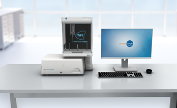
Micro-Flow Imaging (MFI™) gives you more insight into the world of sub-visible particles, going beyond just particle size and count.
With this image-based technology, you can add quantifiable morphological parameters to your analysis, so you can differentiate particle populations from each other and classify them. MFI easily detects translucent protein aggregates that other technologies can miss. Plus, it has an optic focal length that spans the entire fluidic path, giving you robust and reproducible sample data. MFI View System Software and Image Analysis provide 21 CFR Part 11 tools for method and batch execution, data processing and audit trails.
Increase your throughout by adding a Bot1 Autosampler to automate sample processing. If you're looking for broader, deeper analysis of your biotherapeutic products, MFI is the particle analysis tool for you.
Fill out the registration form to start exploring the virtual demo for our Micro-Flow Imaging (MFI) and Bot1 Autosampler.
Before you hit submit, here's Adrian with a brief video to introduce you to MFI. During the virtual demo, you and Adrian will explore these key elements:
© 2023 Bio-Techne. All rights reserved.
Terms and conditions of Sale | Privacy Policy | Email Preferences